
Array-based comparative genomic hybridization (aCGH) is an advanced technique that provides more detailed and comprehensive chromosomal screening, as it analyzes all chromosomes, including the sex chromosomes. Consequently, the selection of embryos with a normal chromosomal complement—free of duplications or deletions—is more accurate and reliable.
For further information or Booking..


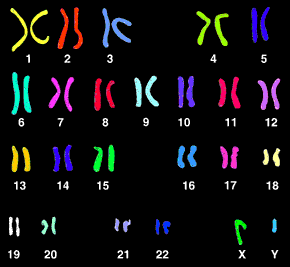
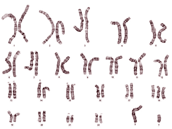
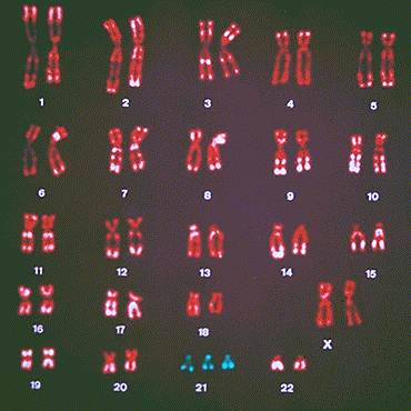
Human Chromosomes
Humans have a total of 23 pairs of chromosomes, consisting of 22 pairs of autosomes (body chromosomes) and 1 pair of sex chromosomes. The XX chromosome combination determines female sex, while XY determines male sex.
What Are Chromosomes?
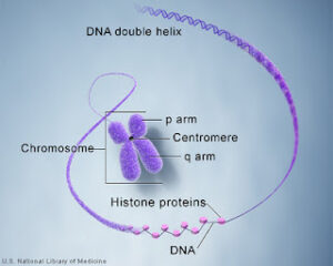
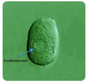
Chromosomes are structures that carry genetic information and are located within the nucleus of cells, the smallest functional units of body tissues. They occur in homologous pairs and are often described as having a shape similar to a pair of pants. Their primary role is to regulate cellular functions, which in turn control how the body’s organs operate.
Each individual’s genetic traits are determined by genes, which are small segments of DNA located on chromosomes. These genes regulate cell growth and function and are responsible for defining the unique characteristics of each person.
Method 1:
Chromosomal Structure
Egg and sperm cells each carry chromosomes, not fragments or components of chromosomes. Each gamete contains half the number of chromosomes required to form a complete genetic set. When the mother’s egg and the father’s sperm unite, their genetic material combines to form a complete set of chromosomes in the embryo. This results in homologous pairs of chromosomes, with one chromosome inherited from each parent.
These genetic codes determine an individual’s unique traits. Although siblings share the same biological parents, they inherit different combinations of genes, which is why each person is unique—even siblings born to the same parents.
Prenatal Genetic Testing Methods
Several methods are currently available for prenatal genetic testing.
Method 1: FISH Technique (Preimplantation Genetic Screening)
This method is now considered outdated and is no longer used for chromosomal testing at Phyathai Sriracha Hospital.
The FISH (Fluorescence In Situ Hybridization) technique detects numerical chromosomal abnormalities by examining the nucleus of a single cell from an embryo. It focuses on identifying common chromosomal disorders observed in newborns, including:
-
Trisomy 13 (Patau syndrome)
-
Trisomy 18 (Edwards syndrome)
-
Trisomy 21 (Down syndrome)
It also screens for abnormalities of the sex chromosomes, such as:
-
XO (Turner syndrome): A condition in which one sex chromosome is missing, resulting in a single X chromosome.
-
XXY (Klinefelter syndrome): Typically presents as male and may be associated with developmental delays and infertility due to impaired or absent sperm production.
Most chromosomal abnormalities occur as a result of errors during egg cell development, which are more common in women over the age of 35.


Method 2: Preimplantation Genetic Testing for Monogenic Disorders (PGT-M)
This method involves screening embryos for specific inherited genetic disorders before implantation. Only embryos without the targeted genetic mutation are selected for transfer, while embryos found to carry the mutation are not transferred.
A common example is thalassemia, a genetic blood disorder in which both parents may be carriers without showing any symptoms. However, their child may inherit the defective gene from both parents and develop the disease. Preimplantation genetic testing helps prevent the transmission of such inherited disorders to offspring.
Advances in genetic testing technology have replaced older techniques, such as FISH (Fluorescence In Situ Hybridization), with more accurate and comprehensive methods, including array Comparative Genomic Hybridization (aCGH).
Method 3: Array Comparative Genomic Hybridization (aCGH) for Higher Accuracy
In the aCGH technique, embryos are first created through in vitro fertilization (IVF). Ovarian stimulation is performed using injectable medications to induce the development of multiple mature eggs, which are then fertilized with sperm in the laboratory.
After fertilization, the embryos are cultured until they reach the appropriate developmental stage. A small number of cells are biopsied from each embryo—typically at the cleavage stage (6–8 cells) or at the blastocyst stage (day 5 after fertilization). These cells are then analyzed using the aCGH technique to detect chromosomal abnormalities across all 23 pairs of chromosomes.
Only embryos with a normal chromosomal profile are selected for transfer to the uterus. This approach increases the likelihood of a healthy pregnancy and reduces the risk of miscarriage and congenital disorders.
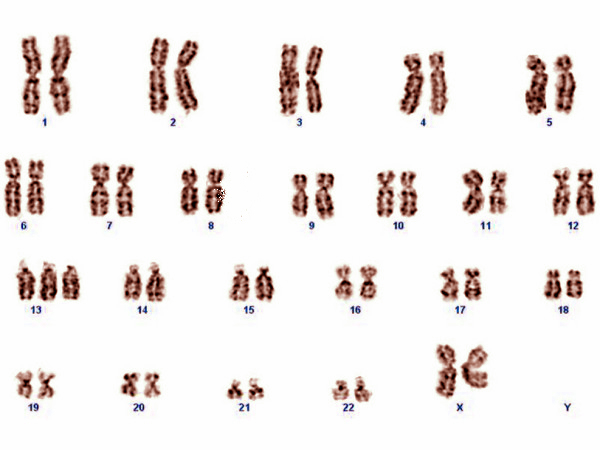
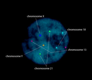
In biopsied embryos, the cytoplasm is removed, leaving only the nuclei. These nuclei are then stained using the FISH (Fluorescence In Situ Hybridization) technique to identify and count specific chromosome pairs, particularly chromosomes 13, 18, 21, X, and Y.
Chromosomal Testing Using the FISH Technique and Its Limitations
Image Interpretation
The image displays the nuclei of the examined embryos, showing two copies each of chromosomes 13, 18, and 21. The presence of XY sex chromosomes indicates a male embryo.
Advantages and Disadvantages of Chromosomal Testing Using the FISH Technique
In women over the age of 35, the risk of chromosomal abnormalities increases due to potential errors during egg development. When abnormal eggs are fertilized by sperm, the resulting embryos may carry chromosomal abnormalities, which can prevent implantation or lead to miscarriage during the first trimester.
Performing chromosomal testing on embryos prior to uterine transfer helps screen for common chromosomal abnormalities observed in newborns and may reduce the risk of early miscarriage in high-risk patients.
However, retrospective studies comparing outcomes between patients who underwent chromosomal testing and those who did not have demonstrated a lower pregnancy rate in the tested group. This reduction is likely attributable to potential embryo damage during the cell biopsy process, which may result in the loss of viable embryos. In addition, false-positive results associated with the FISH technique may lead to the unnecessary exclusion of chromosomally normal embryos, further reducing the likelihood of successful pregnancy.
Therefore, to maximize the benefits of chromosomal testing, it should be reserved for high-risk cases, such as women over the age of 38 or individuals with a family history of chromosomal disorders, including Down syndrome.
Follow-up After Conception
Even after pregnancy is achieved, amniocentesis remains an important diagnostic tool—particularly for older mothers. While preimplantation screening tests assess only a limited number of chromosomal abnormalities, amniocentesis, typically performed between 16 and 18 weeks of gestation, can detect abnormalities across all chromosomes.
Summary: Benefits and Drawbacks of Chromosomal Screening
Benefits
-
Reduces the risk of transferring embryos with chromosomal abnormalities in high-risk patients
-
Can assist in determining fetal sex in selected cases
Drawbacks
-
Possibility of false-positive results
-
Potential reduction in pregnancy rates due to embryo manipulation and cell biopsy
Advanced Method: Array Comparative Genomic Hybridization (aCGH)
Phyathai Sriracha Hospital now utilizes an advanced method known as array Comparative Genomic Hybridization (aCGH) to overcome the limitations of traditional techniques.
This method enables comprehensive screening of all 23 pairs of chromosomes, including the sex chromosomes, with improved accuracy in identifying embryos with a normal chromosomal profile.
The principle of aCGH involves amplifying the embryo’s genetic material to generate hundreds of thousands of DNA copies. These amplified DNA fragments are then analyzed and compared with a known normal DNA reference to identify any missing or extra chromosomal segments.
Unlike the older FISH technique—which relies on counting fluorescent signals within the nucleus and is susceptible to interpretation errors due to signal fading or overlap—aCGH uses DNA microarray technology. The amplified DNA is hybridized onto a microarray containing reference DNA. If the signal pattern matches the reference, the embryo is confirmed to have a normal chromosomal structure; any deviation indicates a gain or loss of genetic material.
This advanced technique significantly improves the accuracy and reliability of embryo selection, thereby increasing the likelihood of successful implantation and healthy pregnancy outcomes.
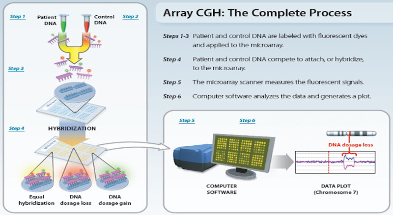 Array CGH: The Complete Process
Steps of Array CGH (Comparative Genomic Hybridization)
Steps 1 & 2
DNA samples are collected from both the embryo (or fetus) and a known reference DNA sample. These samples are analyzed simultaneously, with the reference DNA serving as the control.
Step 3
Both DNA samples are labeled with fluorescent dyes—typically one color for the fetal DNA and a different color for the reference DNA. This labeling allows the two DNA sources to be visually distinguished during analysis.
Step 4
The labeled DNA samples are mixed and hybridized onto a microarray chip containing thousands of known DNA sequences. During this process, the fetal DNA and reference DNA compete to bind to the sequences on the microarray.
Steps 5 & 6
After hybridization, the microarray is scanned using a high-resolution fluorescence scanner. The intensity of the fluorescent signals is measured and analyzed using specialized computer software.
-
Increased signal intensity from the fetal DNA compared with the reference DNA indicates a chromosomal gain (duplication) in that region.
-
Decreased signal intensity indicates a chromosomal loss (deletion).
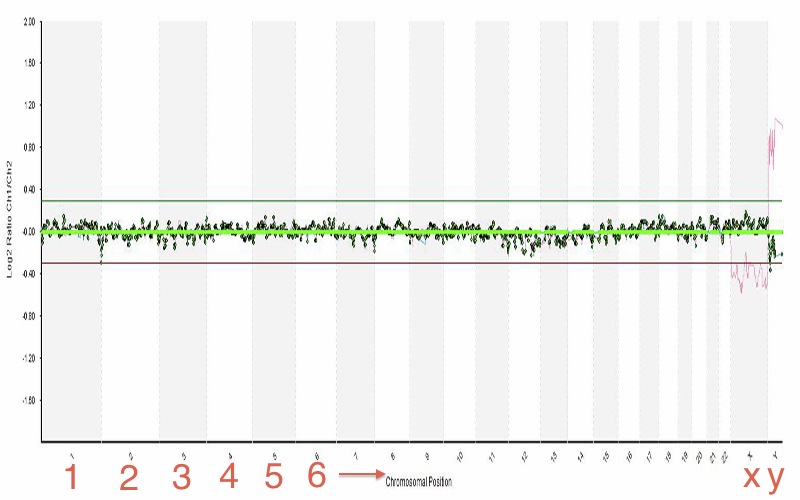
The images present the fetal genetic profile across chromosome pairs 1 through 23. No segments exceed the upper green threshold, which would indicate chromosomal gain, nor fall below the lower red threshold, which would indicate chromosomal loss. These findings are consistent with a normal chromosomal pattern.
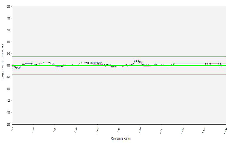
The magnified view of chromosome pair 1 allows for more precise examination of chromosomal details compared with standard paired views, in which chromosomes typically appear as condensed segments. This magnification enables the detection of small duplications or deletions at specific chromosomal loci.
The accompanying video graphically illustrates the fetal chromosomal profile across chromosome pairs 1 through 23. In a normal result, no segments exceed the upper boundary indicated by the green line (representing chromosomal gain) or fall below the lower boundary indicated by the red line (representing chromosomal loss). In addition, each chromosome pair can be individually enlarged and examined in greater detail, allowing for clearer and more accurate analysis than conventional chromosomal overviews.
For further information or Booking..


